SEARCH THIS WEBSITE
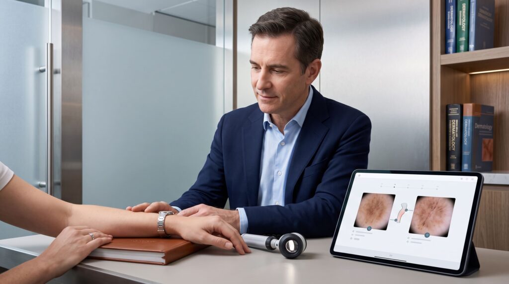
Did you know that seborrhoeic keratoses are among the most common benign skin growths, yet they can closely mimic melanoma, one of the most serious forms of skin cancer? Distinguishing the two is a frequent challenge in primary care, where visual assessment alone can sometimes miss rare but important cases. A Dermatologist with dermoscopy offers the most reliable way to differentiate them and decide whether a biopsy is needed.
Key Areas We Will Cover
- What seborrhoeic keratosis is and how it typically appears
- Key visual differences from melanoma
- Why primary-care diagnosis can be challenging
- The role of dermoscopy and specialist assessment
- When a biopsy is recommended
- Practical advice for UK patients
Introduction
Seborrhoeic keratosis (also called seborrhoeic wart) is a harmless, age-related skin growth that becomes increasingly common after middle age. Melanoma, by contrast, is a potentially life-threatening skin cancer that arises from pigment-producing cells. Because some melanomas can resemble seborrhoeic keratoses, and inflamed seborrhoeic keratoses can look worrying, accurate distinction is essential. Recent commentary aimed at primary-care clinicians highlights simple visual clues while stressing that uncertainty should always prompt further investigation. This article explains the differences and why assessment by a Dermatologist remains the safest approach. At The Skin Care Network our consultant Dermatologists regularly evaluate these lesions using clinical examination and dermoscopy.
What Is a Seborrhoeic Keratosis?
A seborrhoeic keratosis is a benign growth of surface skin cells that typically appears in middle-aged and older adults.
These lesions often have a distinctive “stuck-on” appearance, as if they were glued onto the skin. They may feel waxy, crumbly or crusty and can range in colour from light tan to dark brown or almost black. Most remain stable for years and cause no symptoms, although they can occasionally become irritated or itchy. They commonly appear on the trunk, face and scalp and frequently occur in multiples.
How Does Melanoma Differ in Appearance?
Melanoma tends to show irregular features and change over time.
Warning signs include asymmetry, uneven or blurred borders, multiple colours within the same lesion (brown, black, red, blue or white), larger size, and any evolution in shape, colour or symptoms such as bleeding or itching. The “ugly duckling” sign a lesion that stands out from the person’s other moles or growths is another important clue. While many melanomas are flat or slightly raised, some can develop a rough surface that superficially resembles a seborrhoeic keratosis.
Why Can These Lesions Be Difficult to Distinguish in Primary Care?
Visual overlap and limited access to specialised tools create diagnostic challenges.
Studies have shown that a small proportion of lesions clinically diagnosed as seborrhoeic keratosis (around 0.7 % in one large series) later prove to be melanoma on histology. Inflamed seborrhoeic keratoses are particularly deceptive and can closely mimic melanoma. Primary-care clinicians often work without a dermatoscope and see a high volume of lesions, so the safest approach when any doubt exists is referral or biopsy rather than simple reassurance.
How Does a Dermatologist Improve Diagnostic Accuracy?
Specialist assessment combines experience with dermoscopy.
A Dermatologist examines the lesion under magnification with a dermatoscope, revealing subsurface structures invisible to the naked eye. Classic dermoscopic features of seborrhoeic keratosis include milia-like cysts, comedo-like openings, fissures and ridges, and a sharp border. Melanoma more often shows atypical pigment networks, irregular dots or globules, blue-white veil or other concerning patterns. When these tools still leave uncertainty, a biopsy provides definitive histological confirmation.
When Is a Biopsy Recommended?
Any lesion that cannot be confidently diagnosed as benign should be sampled.
Particular triggers for biopsy include rapid change, irregular colour or border, symptoms such as bleeding or itching, an “ugly duckling” appearance, or simply persistent diagnostic doubt. An inflamed seborrhoeic keratosis is a classic example where biopsy is often the prudent choice. The procedure is quick, performed under local anaesthetic, and allows precise diagnosis so that appropriate treatment (or reassurance) can follow.
What Should UK Patients Do If They Notice a Concerning Lesion?
Prompt specialist review offers the greatest peace of mind.
Perform regular self-checks of your skin and note any new growths or changes in existing ones. If a lesion looks stuck-on and matches the typical description of seborrhoeic keratosis, it is usually harmless, but any uncertainty, change or worry warrants professional assessment.
At The Skin Care Network, located at 68-70 Union St, Barnet EN5 4HZ, United Kingdom, our consultant Dermatologists provide expert evaluation of pigmented and non-pigmented lesions using clinical examination and dermoscopy. Call +44 20 8441 1043 to book an appointment or discuss any skin concerns.
Conclusion
Seborrhoeic keratoses are common, benign and usually easy to recognise by their stuck-on, uniform appearance. Melanoma is far less common but can occasionally mimic these growths, and inflamed seborrhoeic keratoses can look alarming. Primary-care assessment has inherent limitations, which is why specialist review by a Dermatologist with dermoscopy and biopsy when needed remains the most reliable way to distinguish the two and ensure nothing important is missed. Early specialist input supports both accurate diagnosis and appropriate management.
Get Started Today
Unsure whether a skin growth is a seborrhoeic keratosis or something that needs further checks? Book a consultation with a Dermatologist at The Skin Care Network for expert assessment and clear advice.
Frequently Asked Questions
This FAQ section answers common questions about seborrhoeic keratosis and melanoma, helping you make informed decisions with guidance from a Dermatologist.
No. Seborrhoeic keratoses are benign and do not transform into melanoma. However, a melanoma can occasionally look similar, which is why uncertain lesions should be properly assessed.
Experienced clinicians can often recognise classic seborrhoeic keratoses, but studies show that a small percentage of lesions thought to be seborrhoeic keratosis turn out to be melanoma. Dermoscopy and, when needed, biopsy significantly improve accuracy.
The inflammation itself is not dangerous, but the appearance can closely resemble melanoma. Biopsy is frequently recommended in these cases to confirm the diagnosis.
Most do not. Treatment is optional and usually considered only if the lesion is irritated, itchy or cosmetically bothersome. Removal options include cryotherapy, curettage or shave excision after the diagnosis is secure.
If a lesion is changing, looks different from your other growths, causes symptoms, or simply worries you, specialist assessment is advisable. A Dermatologist can provide dermoscopy and arrange biopsy if required, often in a single visit.